PhD defense by Mads Møller Pedersen, MD and PhD student at Rigshospitalet and Center for Fast Ultrasound Imaging
 Mads Møller Pedersen will be defending his thesis on 2 July 2012 at 2 pm.
The defense will take place at Rigshospitalet, Auditorium 93, Julie Mariesvej 93, 2100 København Ø. Check map
Abstract:
This PhD thesis has investigated the use of a new ultrasound technique that to measure the movement of blood. The technique was developed at the Center for Fast Ultrasound Imaging at the Technical University of Denmark and has previously only been available
with experimental ultrasound scanners. Now, the method has been implemented into a commercial ultrasound scanner made for hospital use.
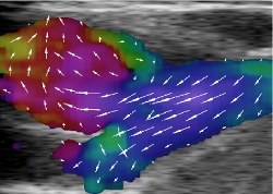
In real-time, the technique measures movements in all directions as 2D vector fields, including movements perpendicular to the ultrasound beam. This is not available with conventional ultrasound scanners today.
The thesis consists of three studies that uses vector flow ultrasound measurements on healthy volunteers. In study I the common carotid artery of 16 healthy volunteers were scanned simultaneously with the vector technique and the conventional, spectral estimation method. The study compared the clinical parameters: peak systole velocity, end diastole velocity, resistive index, and the flow direction. The results showed significant difference on the velocities and the resistive index. However, no significant difference on the manually defined flow angle and the calculated mean flow angle by the vector technique. With the conventional technique, the manual setting of the angle is operator dependent. With the calculated vector angle, this operator is relieved from the angle setting and the measurement is angle corrected by the identical method every time.
With study II the carotid bifurcation including the carotid bulb and the common carotid artery were scanned on 8 healthy volunteers. The flow patterns of the two structures were outlined and presented to each of 5 experienced radiologists. The complexity of the identical areas were calculated by the vector concentration and compared to the visual evaluations. No significant difference was found between the two methods which were equally good at discriminating the laminar flow of the common carotid artery from the complex flow in the carotid bulb. Thus, a new method was presented to quantify complex flow patterns with vector flow.
The final study III presented the rotational flow patterns in the cross-sectional plane of three arteries: The common carotid artery, the abdominal aorta, and the common iliac artery. Five healthy volunteers were included in the study and nine datasets visualized the flow patterns during the diastole. The rotational frequency was calculated and the results indicate a constant direction of the rotation for each artery. Extended measurements on the abdominal aorta showed a two-directional rotation during the cardiac cycle. An observation that corresponds to previous MR and Doppler studies.
With the three studies, this thesis presents new methods that quantifies in vivo vector flow obtained in real-time with a new implementation.
Download thesis